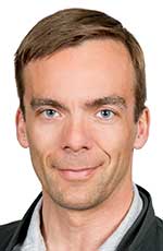
Futuristic handheld scanners that can instantly diagnose all that ails us are on the verge of becoming a reality, thanks to a University of Alberta researcher whose use of artificial intelligence (AI) to pinpoint a growing host of disorders landed him an unprecedented research chair.
Jacob Jaremko, a pediatric and musculoskeletal radiologist and researcher in the Faculty of Medicine & Dentistry, has become the only practising physician to be awarded a Canadian Institute for Advanced Research (CIFAR) AI Chair. He has been granted this chair at the Alberta Machine Intelligence Institute (Amii) for his application of artificial intelligence to medical problems.
“As a clinician-scientist I kind of straddle that divide,” said Jaremko, who completed a PhD in biomedical engineering and developed expertise in the earliest form of AI before attending medical school at the U of A. “I hold a unique position in that way.”
Jaremko’s team is particularly interested in the use of portable ultrasound machines alongside AI to play a larger role in diagnosing injury and illness.
In his most recent study, his team looked at whether a quick scan from an ultrasound machine could be as effective as an X-ray in diagnosing broken arms in children visiting an emergency room.
The current process of waiting to see a physician, going for X-rays and getting a diagnosis takes several hours. As well, roughly half of the time there is no fracture.
Ultrasound can be instantaneous, but the black-and-white snowstorm images can be quite confusing for the uninitiated to interpret, noted Jaremko, who holds the Alberta Health Services Endowed Chair in Diagnostic Imaging at the U of A and is a member of the Women’s and Children’s Health Research Institute.
“However, an artificial intelligence network can recognize patterns in the ultrasound, much the same way that your iPhone can recognize your face and unlock itself.”
For the study, a cohort of ultrasound experts reviewed both ultrasound and X-ray images of bone breaks. They found that, with the right expertise, ultrasounds were as good as X-rays in identifying fractures.
Those same images were then fed into a neural network that researchers in Jaremko’s lab trained to search for fractures. The study showed the neural network accuracy was the same as the human expert accuracy.
“In the future, a child with a sore arm comes in and instead of sending them for this several-hours-long procedure, (the clinician) scans the patient for a few seconds, looks at the pictures and gets an opinion from the computer as to whether a fracture is detected,” he said.
“In at least half of the kids there is no fracture, so you’re done – they go home and an emergency room bed opens up. Similarly, you could expand that out to walk-in clinics, ambulances and remote communities.”
Jaremko said this “21st-century stethoscope” doesn’t just stop at fractures but can also be used to look into the body and detect many kinds of pathology.
In 2017, Jaremko, former U of A post-doctoral fellow Dornoosh Zonoobi and Jeevesh Kapur, a radiologist from Singapore, with support from Alberta Innovates, formed U of A spinoff MEDO.ai, a startup based on the artificial intelligence analysis of images.
MEDO is in the process of commercializing a pair of ultrasound technologies developed by Jaremko’s lab, both of which are already FDA approved. The first revolves around the prenatal diagnosis of hip dysplasia, which occurs when the ball and socket joint is poorly formed.
Though this affliction is easy to correct at the earliest stages of life, it often goes undiagnosed, which dooms those who live with it to a life of pain, suffering and limited mobility.
Using AI, MEDO trained a neural network using ultrasound images of hip dysplasias to reliably detect the disorder. It is currently being piloted in some clinics in the Edmonton region, the U.K. and Brazil.
Another technology will help physicians identify types of thyroid nodules.
“Here’s something you could do in the clinic, scan more patients and reduce wait times,” he said.
“Especially in follow-up, if you’re always doing the scan with the same computer system, then you get a very consistent approach and you can say whether the nodules changed. The computer network really supports thyroid ultrasound and makes it a lot more robust and a lot easier to use.”
| By Michael Brown
This article was submitted by the University of Alberta’s Folio online magazine. The University of Alberta is a Troy Media Editorial Content Provider Partner.
© Troy Media
Troy Media is an editorial content provider to media outlets and its own hosted community news outlets across Canada.


