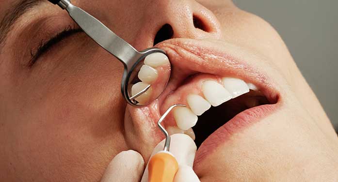
Dentists may soon add portable ultrasound to the standard equipment in their offices, allowing them to accurately and affordably diagnose aspects of their patients’ periodontal disease without any risk of radiation.
That’s the goal of a University of Alberta research team that will continue to develop and commercialize its patented dental ultrasound system thanks to a new grant from Alberta Innovates.
 Lawrence Le |
 Paul Major |
The team has already developed a handheld, portable ultrasound device that can easily and comfortably be used to produce two-dimensional images inside the mouth. The new funding will support the next step in development – producing reliable three-dimensional images to aid further in the detection of dental disease.
“Having a 2D image is like being in the forest with no bearings and no idea what’s going on in the neighbourhood,” says principal investigator Lawrence Le, clinical professor in the Department of Radiology and Diagnostic Imaging. “By transforming 2D images into 3D images, we will be able to really look around at different angles, giving a view of the soft tissue, blood flow and bone.”
The 3D ultrasound will eventually be tested in clinical trials to diagnose periodontal gum disease, which affects up to 90 per cent of the population and can lead to tooth loss if left untreated. The researchers foresee that the system could one day be used to guide dental implant design, monitor oral lesions and even diagnose cavities.
Currently, the only 3D imaging tool available to thoroughly assess periodontal gum disease is cone-beam computed tomography (CBCT), which rotates around the patient’s head using a cone-shaped X-ray beam. It’s a large, expensive machine usually only available in specialists’ offices. CBCT exposes patients to high levels of radiation, so it can’t be used routinely on children or to track changes over time with repeat scans.
Ultrasound is more portable and more affordable and is already widely used in medical settings. In dentistry, ultrasound technology has mainly been used for teeth cleaning rather than imaging. The U of A research team began working together eight years ago to change that.
| RELATED CONTENT |
| Five ways to keep your smile healthy By Gillian Anderson |
| Making health care more equitable one ultrasound image at a time By Gillian Anderson |
| Ultrasound has potential for treating pain after chemotherapy By Adrianna MacPherson |
Paul Major, an orthodontist, professor and chair of the School of Dentistry, was looking for a way to examine the position of bone in relation to overcrowded teeth to determine the best course of treatment. He turned to Le in radiology for help.
“We started by using a medical ultrasound unit, but, of course, the probe is big and doesn’t really fit well in the mouth, so the problem became how we miniaturize this imaging tool such that it’s easily used inside the mouth. There was nothing commercially available, so we started there,” says Major.
A U of A spinoff company, DenSonics Imaging, has been set up to commercialize the ultrasound device, which is being clinically tested for reliability on patients at the School of Dentistry’s Oral Health Clinic in advance of applying for Health Canada approval. Both Le and Major are members of the Women and Children’s Health Research Institute.
The 3D system will use artificial intelligence to assist non-expert operators in dental offices to perform the tests and interpret the results. An external tracker will be developed to constantly recalibrate the position of the probe as it is moved around in the mouth to take the images. Then visualization software will fuse the 2D images into a 3D “movie” that shows the teeth in the context of the bone and soft tissue.
“We’ve done focus groups with various clinicians in private practice, and the 3D visualization was top of the list for most,” says Major. “As dentists, we’re not actually trained in ultrasound imaging assessment, so there’s a bit of a learning curve. Artificial intelligence can help with that.
“Our vision would be that the dentist could interpret the image for the patient’s benefit right there in their office.”
Both researchers agreed the new provincial funds are crucial to hiring staff and supporting graduate students who will develop and test the system. “Without the funding, we definitely would not be able to move forward,” says Le.
| By Gillian Rutherford
Gillian is a reporter with the University of Alberta’s Folio online magazine. The University of Alberta is a Troy Media Editorial Content Provider Partner.
The opinions expressed by our columnists and contributors are theirs alone and do not inherently or expressly reflect the views of our publication.
© Troy Media
Troy Media is an editorial content provider to media outlets and its own hosted community news outlets across Canada.


